
Retinal Detachment and Retinal Tears
A retinal tear and a retinal detachment are the same problem caught at two different moments. A tear can be sealed with laser in the consulting rooms in a few minutes. Once the retina has detached, treatment usually means a considerably more invasive procedure.
What separates the two is usually nothing more than how quickly the eye was assessed.
New Flashes, Floaters or a Shadow: Call Today
New flashes of light, a sudden increase in floaters, or a shadow anywhere in the vision should be assessed the same day or the next. Call (02) 8544 0719.
Do not wait to see whether the symptoms settle. A tear can progress to a detachment within days, and the symptoms that precede a detachment are the same ones that precede a tear.
Posterior Vitreous Detachment: Where It Starts
The eye is filled with a clear gel called the vitreous, which in youth is attached to the surface of the retina. From around the mid-forties the gel begins to shrink and liquefy, and in time its outer layer peels away. This is a posterior vitreous detachment, and it happens to most people eventually.
On its own it is not a disease and it needs no treatment. It is worth understanding because of what it can do on the way past: as the gel separates, it tugs on the retina, and occasionally it pulls hard enough in one spot to tear it.

Those symptoms are the subject of the flashes and floaters page, which covers what to do when they appear.
From a Tear to a Detachment
A retinal tear is a small break in the retina, usually out in the periphery where the gel was most firmly attached. By itself it does not lift the retina and it does not take the vision.
The trouble is what a tear allows. Fluid inside the eye passes through the break and collects underneath, and the retina begins to lift away from the wall of the eye like wallpaper coming off a damp wall. That is a retinal detachment. As it spreads, the patient sees a shadow or curtain moving in from the edge of the vision, and once it reaches the macula the central vision goes with it.
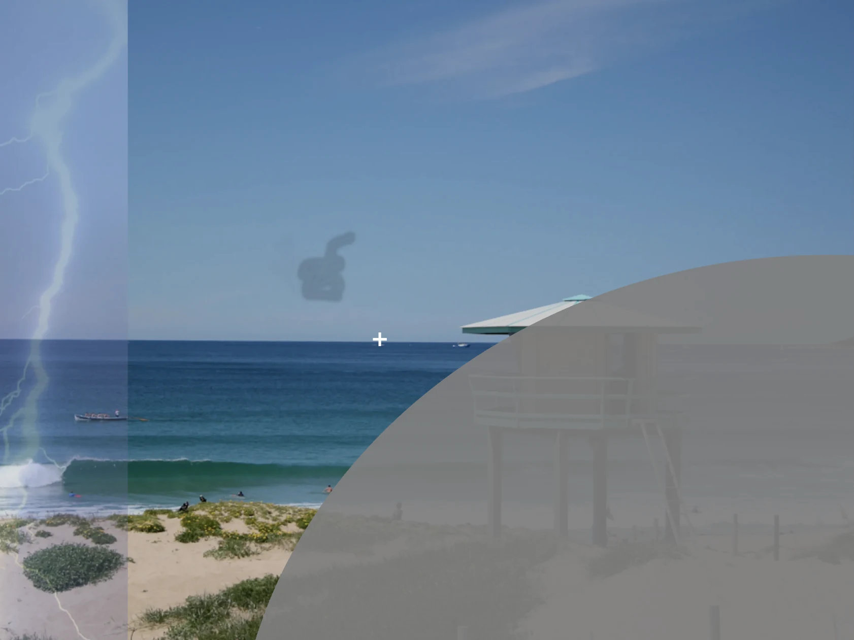
None of this hurts. There is no pain at any stage, which is why the visual symptoms are the only warning available.
Why Early Presentation Changes the Treatment
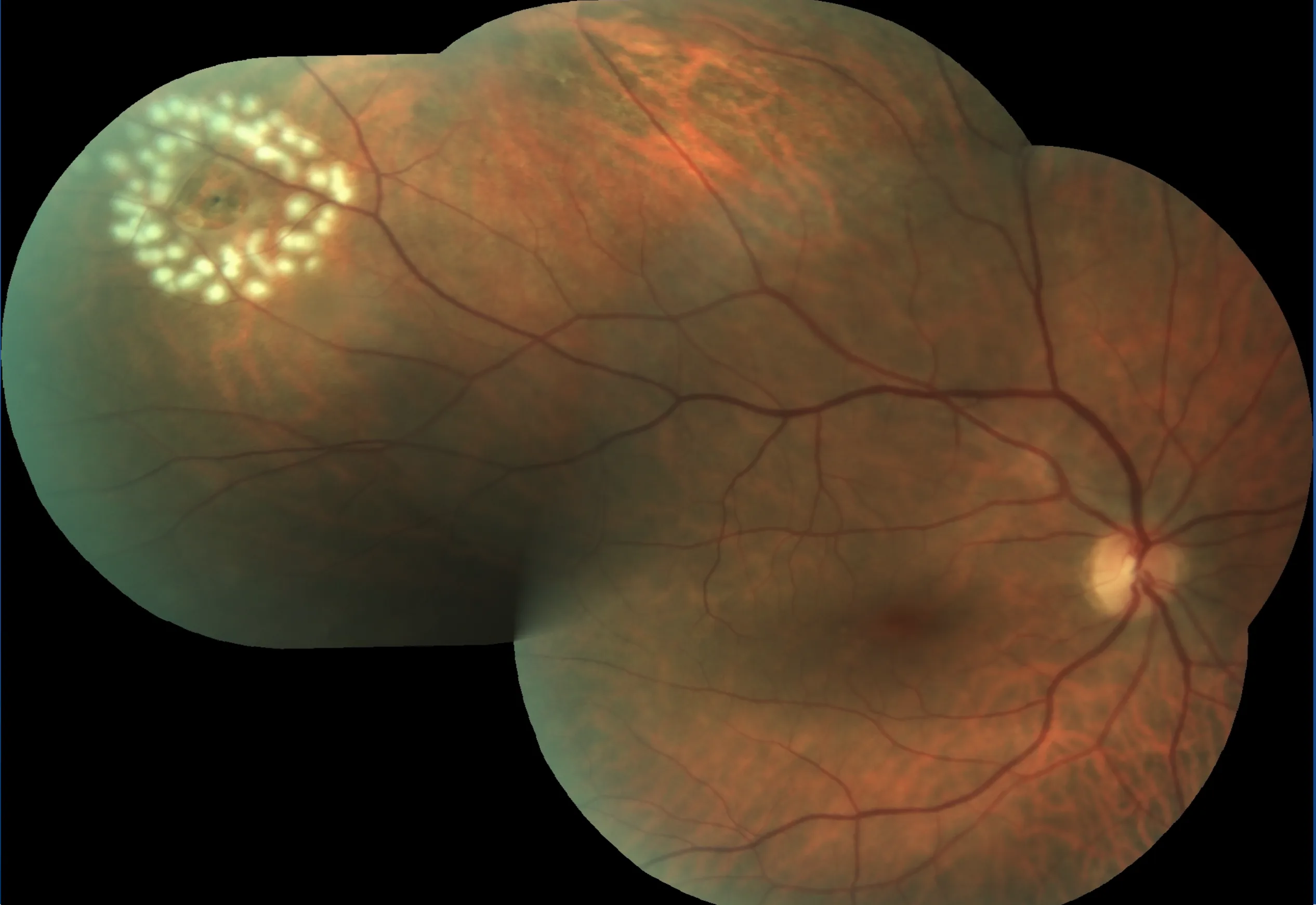
The two illustrations above are what this feels like from the inside. The photographs below are what the same two situations actually look like on examination, and the difference between them is the whole argument for coming in early.
When a tear is found before the retina has detached, it is treated with laser in the consulting rooms. The laser places a ring of small burns around the tear, and as those heal they scar the retina down to the tissue beneath, walling the break off so fluid cannot spread underneath it. The procedure takes minutes, requires no operation, and the seal is permanent once healed.
When the same tear is left long enough for fluid to get underneath, laser can no longer hold it. A detached retina has to be reattached surgically in an operating theatre, with a longer recovery and a result that is less predictable, particularly if the central vision was already involved.
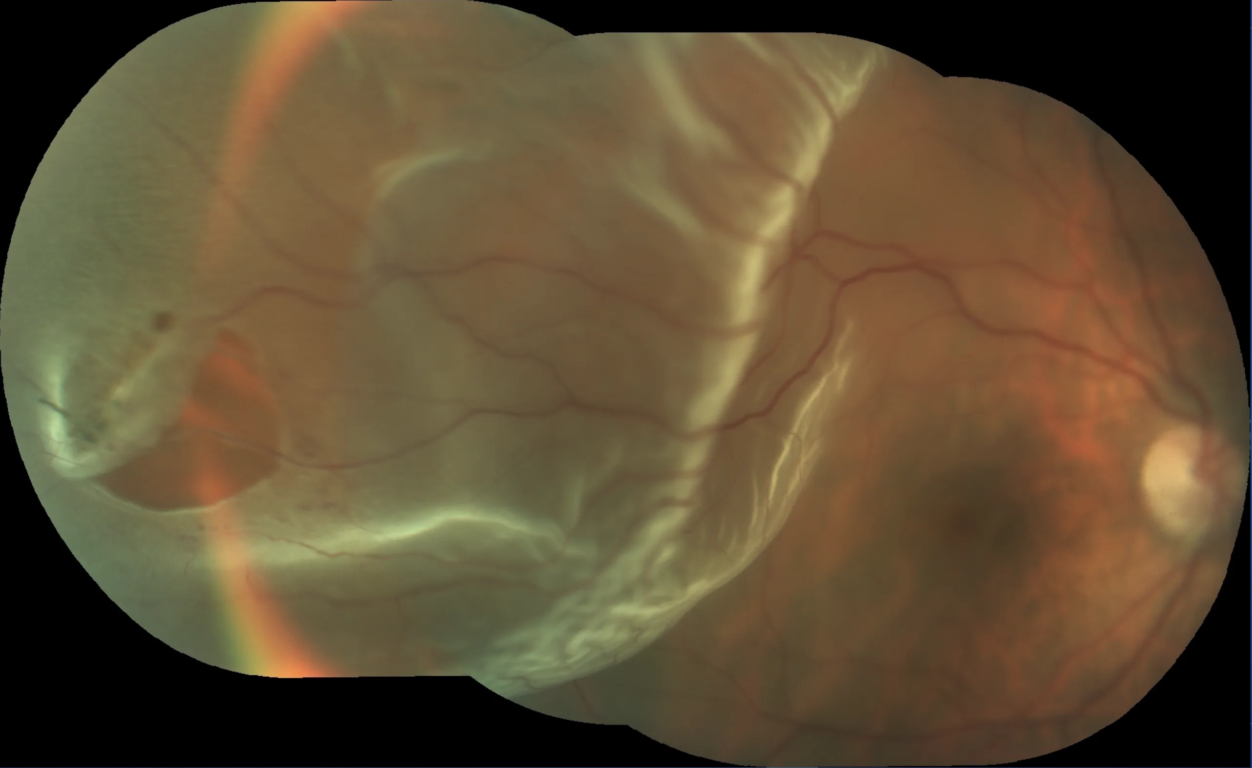
The clinical difference between those two photographs is a matter of timing rather than of luck, and it is the single strongest argument for having new flashes and floaters looked at straight away.
Who Is at Higher Risk
Retinal tears and detachments are more likely in people who:
- Are short-sighted, particularly strongly short-sighted
- Have had cataract surgery
- Have had a tear or detachment in either eye before
- Have a family history of retinal detachment
- Have had a significant injury to the eye
Anyone in these groups who develops new flashes or floaters should be assessed promptly rather than waiting to see what happens. A dilated examination is the only way to inspect the peripheral retina properly, so you will need someone to drive you home.
Frequently Asked Questions
Is a posterior vitreous detachment the same as a retinal detachment?
No, and the difference matters. A posterior vitreous detachment is the gel inside the eye separating from the retina. It is an ordinary age-related change and usually needs no treatment. A retinal detachment is the retina itself lifting away from the wall of the eye, which threatens sight and requires surgery. The first is common and the second is not, but the first is how the second usually begins.
Can a retinal tear be treated without surgery?
Yes, provided it is found before the retina has detached. A tear on its own is sealed with laser in the consulting rooms, in a procedure that takes a few minutes and requires no operation. Once the retina has detached, laser is no longer sufficient and surgery in an operating theatre is required.
Who is at higher risk of a retinal tear or detachment?
Short-sightedness is the most important risk factor, particularly when strong. Others include previous cataract surgery, a previous tear or detachment in either eye, a family history, and significant injury to the eye. Anyone in these groups who develops new flashes or floaters should be assessed without delay.
Does a retinal tear cause pain?
No. Retinal tears and detachments are painless, which is part of what makes them dangerous. There is nothing to warn you other than the visual symptoms: new flashes, new floaters, or a shadow encroaching from the edge of vision.