OCT Imaging — Retinal and Optic Nerve Scanning
Optical coherence tomography (OCT) produces a cross-sectional image of the retina and optic nerve in microscopic detail — far beyond what can be seen with a standard eye examination. It is the single most important diagnostic tool in modern retinal and glaucoma care, and A/Prof Hunt uses it at virtually every retinal and glaucoma consultation to guide diagnosis, monitor disease, and decide when treatment is needed.
On-Site OCT at Eye Surgeons Miranda
The practice is equipped with a Zeiss Cirrus 6000 OCT with AngioPlex — a high-resolution platform that provides both structural imaging of the retina and optic nerve and OCT angiography (OCT-A), which maps the retinal blood vessels without needing a dye injection.
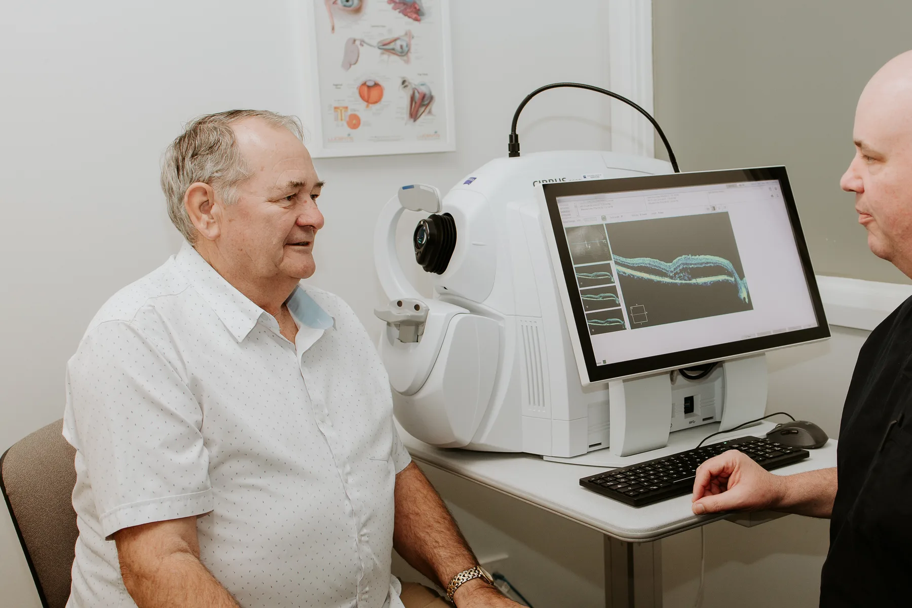
Having OCT on-site means imaging and clinical decisions happen in the same visit. There is no need to travel to a separate imaging centre and return with results — A/Prof Hunt reviews the scan with you at the time and explains what it shows.
What Does an OCT Scan Show?
An OCT scan reveals the individual layers of the retina and the structure of the optic nerve in a way that no other test can. Depending on the condition, it can show:
- Fluid beneath or within the retina — the hallmark of wet macular degeneration, retinal vein occlusion, and diabetic macular oedema. The presence or absence of fluid is what drives injection treatment decisions at every visit.
- Drusen and early structural change — in dry macular degeneration, allowing monitoring for progression toward the wet form.
- Membrane formation on the retinal surface — in epiretinal membrane, showing the degree of wrinkling and traction on the macula.
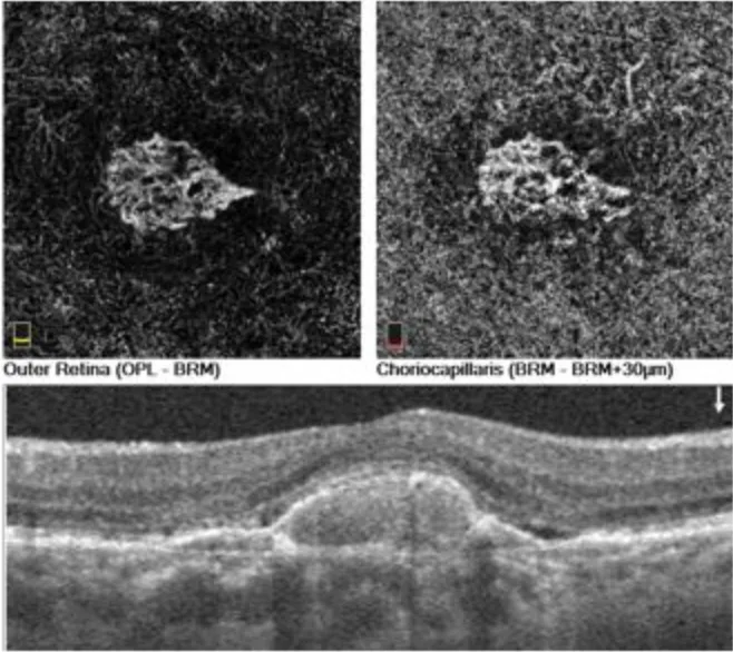
- The structure and thickness of retinal or choroidal lesions — such as naevi (moles) found during routine examination. A cross-sectional image helps characterise the lesion and monitor it for change over time.
- Nerve fibre layer thickness — in glaucoma, where OCT establishes a baseline and tracks change over time to detect progression.
OCT has become an invaluable tool across much of ophthalmology — not only retinal disease and glaucoma. Anterior-segment OCT can image the cornea, anterior chamber angle and iris, and high-resolution OCT is increasingly used in surgical planning and post-operative follow-up, including around epiretinal membrane peeling and retinal detachment repair.
OCT Angiography (OCT-A)
OCT angiography maps blood flow within the retina without injecting any dye. Although the practice has the capacity for traditional intravenous fundus fluorescein angiography, much of that work is now performed non-invasively using OCT angiography — avoiding the intravenous injection of a fluorescent dye and the potential side effects associated with it.
OCT-A detects areas of reduced perfusion, abnormal new vessels, and subtle vascular changes that standard OCT alone may miss. It is particularly useful in diabetic eye disease and retinal vein occlusion, where the pattern of blood flow helps determine disease severity and guide treatment.
What to Expect
An OCT scan is quick, painless, and non-contact — nothing touches the eye. You sit at the instrument, rest your chin on a support, look at a small target light, and the scan is captured in seconds. No drops are needed for the OCT scan itself, although your pupils may be dilated separately as part of a full examination.
A/Prof Hunt reviews the scan with you during the consultation and explains what it shows and what it means for your treatment.
Common Questions
Is an OCT scan painful?
No. OCT is completely painless and non-contact. You sit at the instrument, look at a target, and the scan is captured in seconds. No drops are needed for the scan itself, although your pupils may be dilated separately as part of a full retinal examination.
How often will I need an OCT scan?
This depends on the condition being monitored. Patients receiving injection treatment for wet macular degeneration or retinal vein occlusion are typically scanned at every visit to guide treatment decisions. Patients with stable glaucoma or dry macular degeneration may be scanned every six to twelve months to check for change.
What is OCT angiography?
OCT angiography maps the blood vessels within the retina without needing a dye injection. It detects areas of reduced blood flow, abnormal new vessels, and subtle vascular changes that may not be visible on a standard OCT scan. It is particularly useful in diabetic eye disease and retinal vein occlusion.
Can OCT detect glaucoma?
OCT can show that the nerve fibre layer is thinner than expected compared with age-matched normals, which raises suspicion — but glaucoma is a diagnosis of change. A single scan establishes a baseline; it is the comparison over time that detects progression and confirms the diagnosis. That is why regular OCT monitoring is so important in glaucoma care.