
Diabetic Retinopathy — How Diabetes Affects the Eyes
Diabetes affects every blood vessel in the body, including the delicate network of vessels in the retina. Because the retina is one of the most metabolically active tissues in the body, it is particularly vulnerable. Over time, those vessels can begin to leak, drop out, or trigger the growth of abnormal new vessels that cause further damage. This spectrum of damage to the retina is what we call diabetic retinopathy.
The disease process is gradual, and closely linked to how long you have had diabetes and how well it has been controlled over that time. But one of the most important things to understand is that serious damage can develop silently — vision can be completely normal until suddenly it is not. Regular monitoring is the only way to catch this before it threatens sight.
Almost all the severe vision loss I see from diabetes could have been prevented, in patients who skipped regular review and came in when everything had gone wrong.
What Eye Problems Does Diabetes Cause?
Diabetic retinopathy begins with microscopic changes in the small blood vessels of the retina. Over years, these can gradually build toward vision-threatening problems:
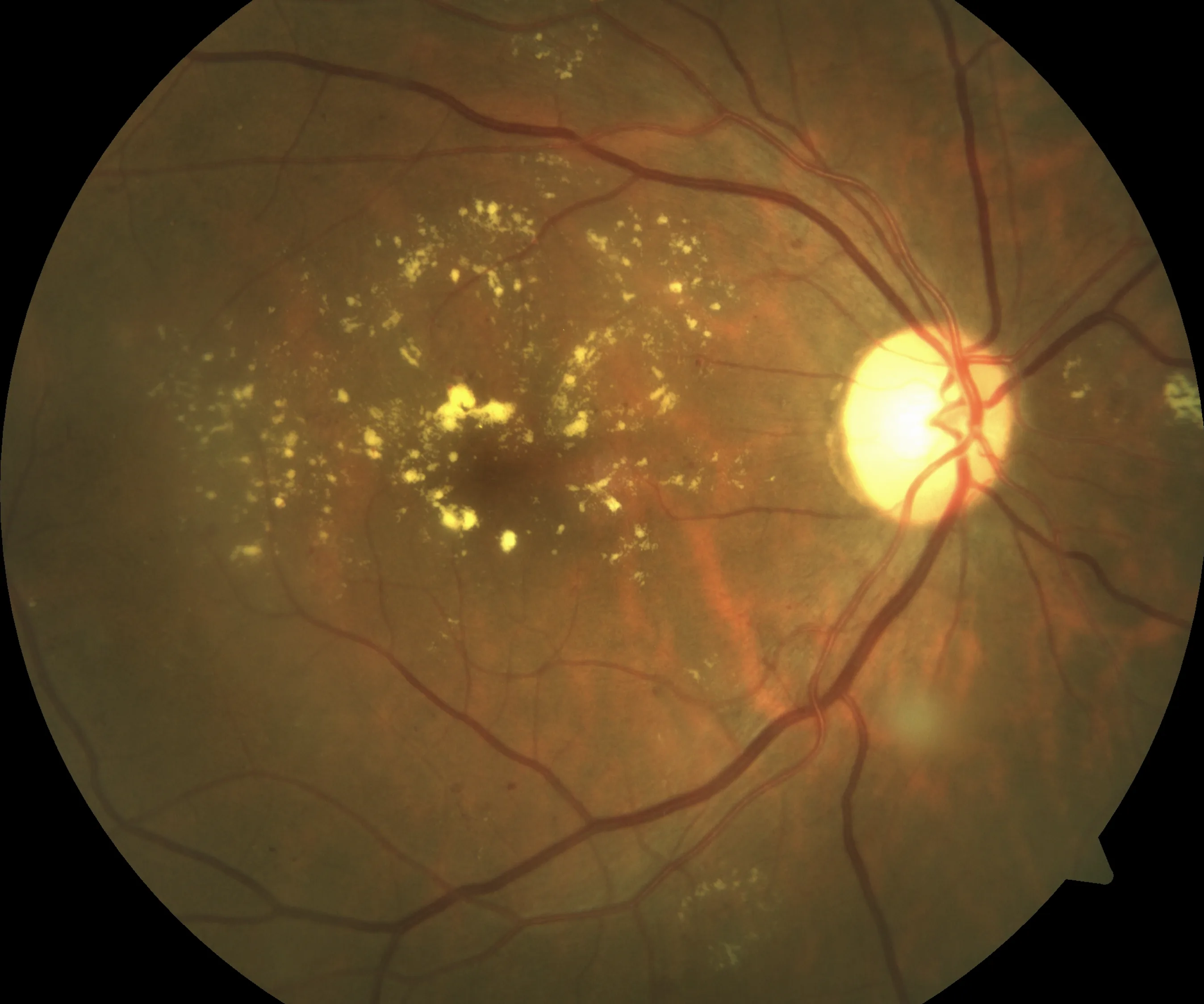
- Diabetic macular oedema (DMO) — damaged vessels leak fluid into the macula, the central part of the retina responsible for detailed vision. The resulting swelling blurs central vision and, if untreated, can cause permanent damage.
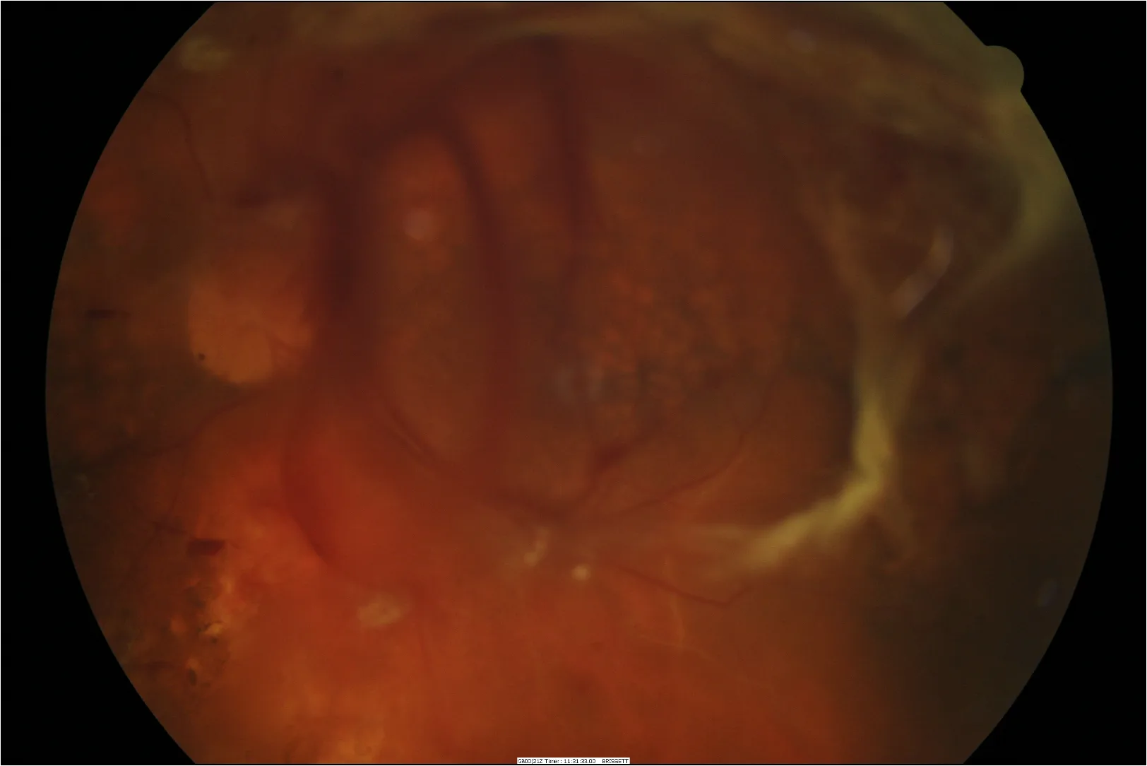
- Proliferative diabetic retinopathy (PDR) — areas of retina that have lost their blood supply become starved of oxygen. In response, the eye grows fragile new vessels that are prone to bleeding into the eye, causing sudden severe vision loss from a vitreous haemorrhage. These vessels can also pull on the retina and cause a tractional retinal detachment.
Both are treatable, particularly when detected early, and the two can develop in the same eye.
Why Diabetic Retinopathy Often Has No Warning Signs
Proliferative diabetic retinopathy — the more advanced and immediately dangerous form — can develop with no symptoms at all. The abnormal new vessels produce no warning signs until they bleed. When that bleeding happens, the vision loss can be sudden and severe.
Many people who lose vision from diabetic retinopathy felt that their eyesight was perfectly normal beforehand. By the time symptoms appear, the retinal damage may already be advanced. Regular diabetic eye examinations are designed to detect problems before they affect vision, when treatment is often simpler and more successful.
Seek Urgent Assessment
A vitreous haemorrhage normally presents with a sudden shower of inky, dark, stringy floaters. This is the typical first sign of a bleed into the back of the eye, which often blurs into a general haze over the following day or so.
Any dark shadow or veil across part of your vision, flashes of light, or other new visual symptoms also need urgent review, as they could indicate a retinal detachment.
For urgent review, call our rooms on (02) 8544 0719 to arrange an urgent appointment. After hours, Sydney Eye Hospital Emergency can provide assessment.
Stages of Diabetic Retinopathy
Diabetic retinopathy is graded by severity, which guides treatment and how often your retina needs to be reviewed.
- No diabetic retinopathy — retinal vessels appear normal; ongoing screening is still required
- Mild non-proliferative — early changes such as microaneurysms only
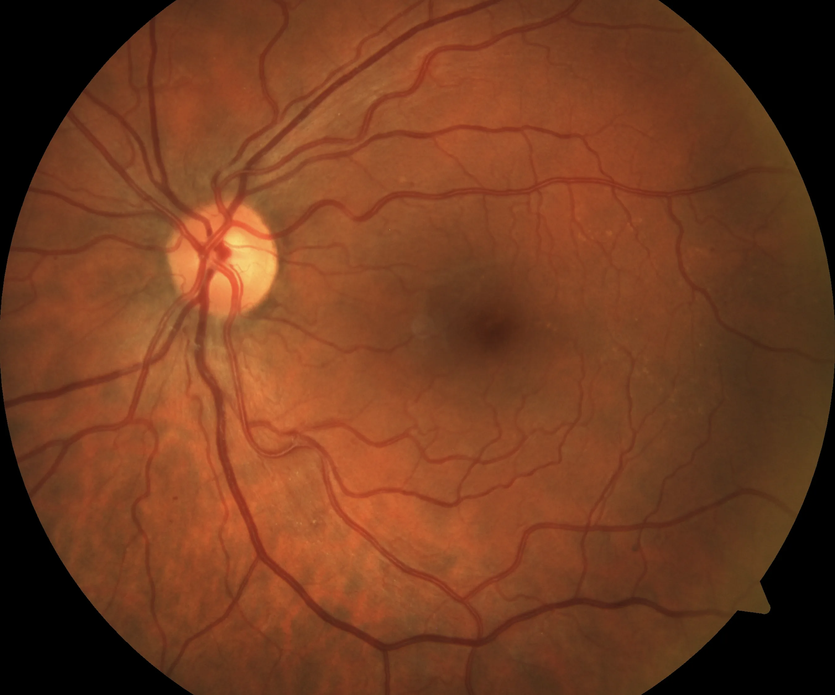
- Moderate non-proliferative — more widespread haemorrhages and exudation, with increasing risk of progression
- Severe non-proliferative — high-risk pre-proliferative disease with a substantial risk of progressing to the proliferative stage
- Proliferative diabetic retinopathy — abnormal new vessels on the retina or optic disc, with risk of bleeding and retinal detachment
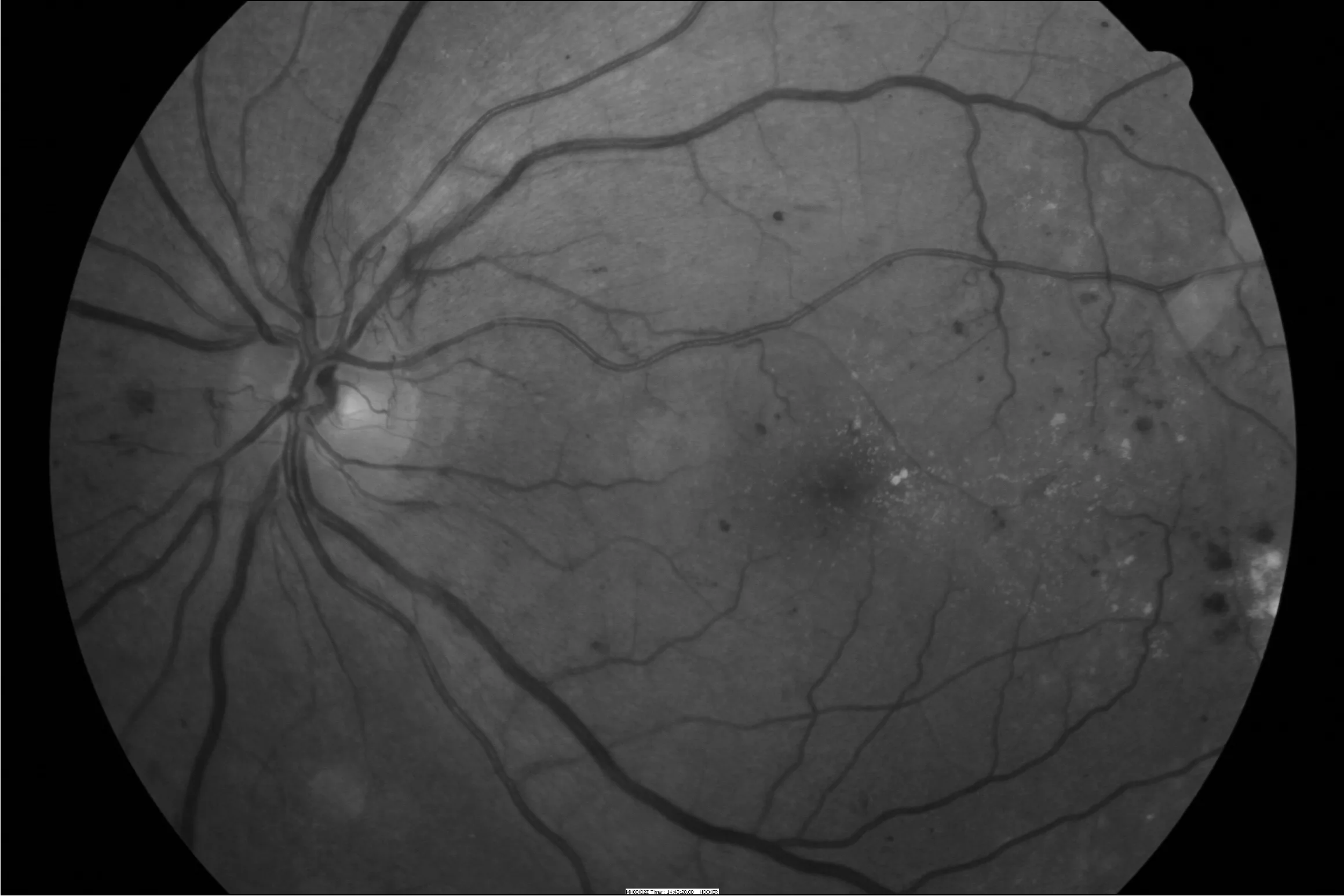
- Diabetic macular oedema — can occur at any stage and is assessed separately from the retinopathy grade
Factors that increase the risk of progression include longer duration of diabetes, poor glycaemic control, high blood pressure, renal disease, and pregnancy.
When Should You Have a Diabetic Eye Check?
Every person with diabetes should have a diabetic eye check — sometimes called diabetic eye screening or retinopathy screening — ideally at or around the time of diagnosis, to establish the starting point of their retinal health. How often you need follow-up depends on what is found at that initial review.
More urgent review is appropriate if you have proliferative changes, centre-involving macular oedema, pre-existing retinopathy in pregnancy, or any sudden change in vision.
Diabetic retinopathy is also a marker of wider blood vessel disease. If retinopathy is found, it is worth discussing with your GP or endocrinologist whether your blood glucose, blood pressure, lipids, and kidney function are all being monitored closely.
Treatment — Injections for Diabetic Macular Oedema
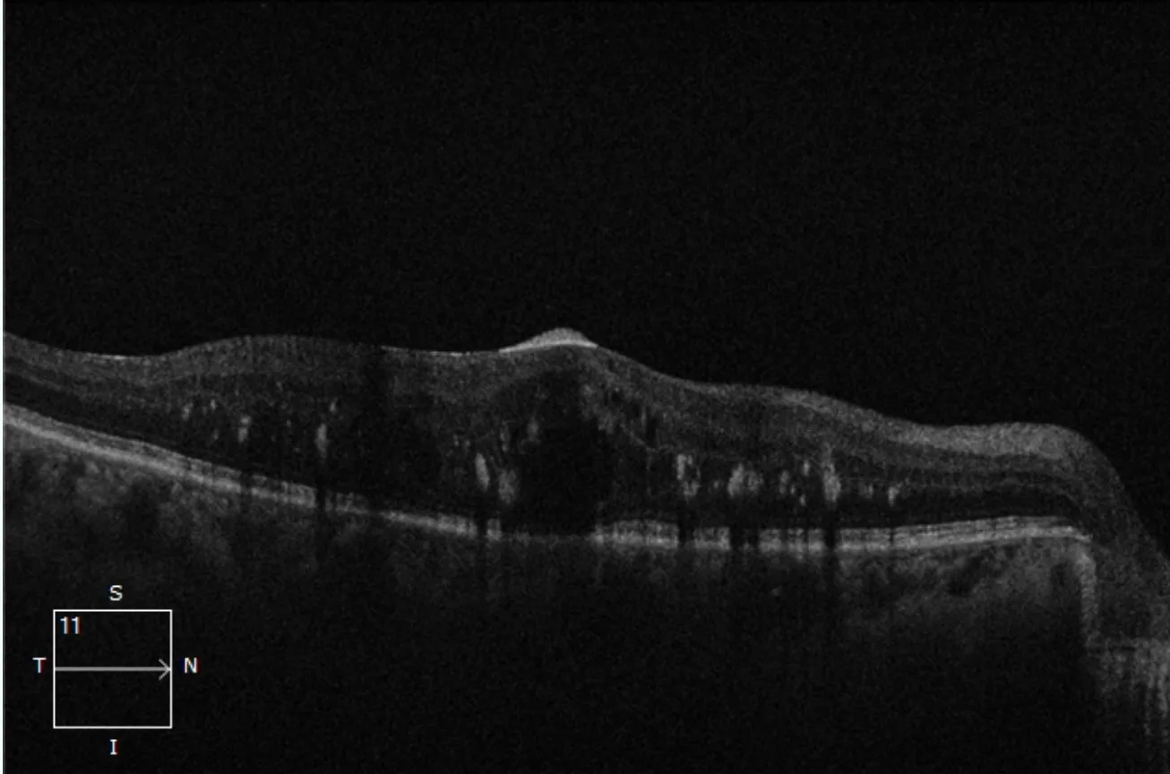
Intravitreal injections are the primary treatment for diabetic macular oedema affecting central vision. Anti-VEGF medications reduce vascular leakage, decrease retinal swelling, and protect the macula. In selected cases — particularly where the response to anti-VEGF therapy is incomplete — a steroid implant may be used instead or in addition.
Treatment response is monitored with OCT imaging at every visit, and the schedule is adjusted accordingly.
Treatment — Laser for Proliferative Diabetic Retinopathy
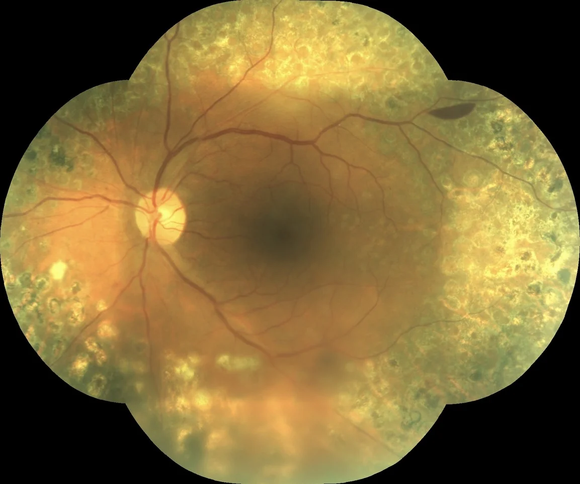
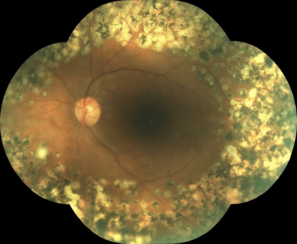
When areas of the retina are starved of oxygen and driving the growth of abnormal new vessels, retinal laser treatment is used to reduce that stimulus. By treating the oxygen-deprived peripheral retina, laser reduces the signal for new vessel growth and protects the eye from bleeding and further complications.
The laser is applied to the oxygen-starved mid-peripheral retina — the tissue driving the abnormal vessels — in order to protect the central retina you rely on for detailed vision. Because this addresses the underlying cause rather than simply suppressing it, the regression of the new vessels tends to be lasting. Occasionally, even after reasonably heavy laser, the neovascular process recurs and further, heavier laser is needed — that is the exception rather than the rule, and it is why the retina is reviewed carefully after treatment.
In practice, injections and laser are often used together — injections can rapidly suppress new vessel activity while a course of laser treatment is being completed, reducing the risk of a vitreous haemorrhage during that period. The important difference is that the injection effect is temporary, whereas the laser provides the more durable foundation.
Research in Diabetic Retinopathy
A/Prof Hunt's involvement with the Fight Retinal Blindness! registry includes research on treatment outcomes in diabetic macular oedema — examining how patients respond over years of therapy, when treatment intervals can be safely extended, and what predicts the best long-term vision. This work directly informs the way diabetic retinopathy is managed in the consulting room.
See A/Prof Hunt's publications →Diabetic Retinopathy Assessment and Treatment
Eye Surgeons Miranda offers baseline assessments for newly diagnosed patients, ongoing retinopathy monitoring, intravitreal injections for diabetic macular oedema, and laser treatment for proliferative disease. Referrals are welcome from GPs, endocrinologists, and optometrists, and urgent cases are seen promptly.